
Figure 5.
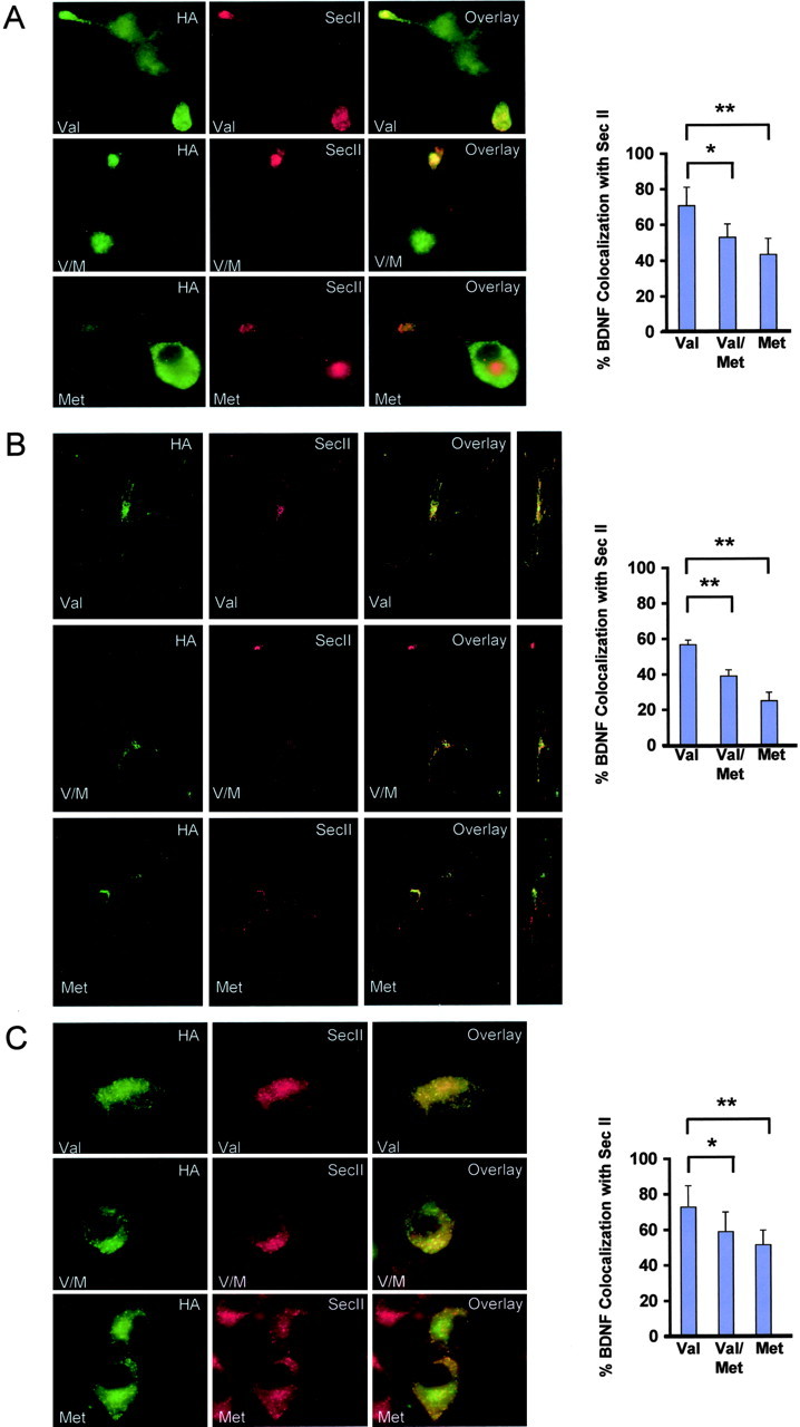
Subcellular colocalization of variant and wild-type BDNF with organelle markers. Differentiated PC12 cells (A), cortical neurons (B), and undifferentiated PC12 cells (C) were transfected with C-terminal HA-tagged variant (Met), C-terminal HA-tagged wild-type BDNF (Val), or both C-terminal FLAG-tagged variant and HA-tagged wild-type BDNF (V/M). Cells were fixed after 48 hr and permeabilized. Colocalization of BDNF and the secretory granule marker SecII was visualized by costaining with anti-HA and anti-SecII antibodies. Confocal three-dimensional reconstruction of serial z planes were obtained, and alternate rotation angle is also shown for each representative cell. Quantitative analysis of results illustrated in A–C was conducted by scoring ∼25 cells for each BDNF condition. The proportion of colocalization between BDNF and SecII is presented as a mean ± SEM determined from analysis of four independent experiments (*p < 0.01; **p < 0.001; Student's t test).