
Figure 3.
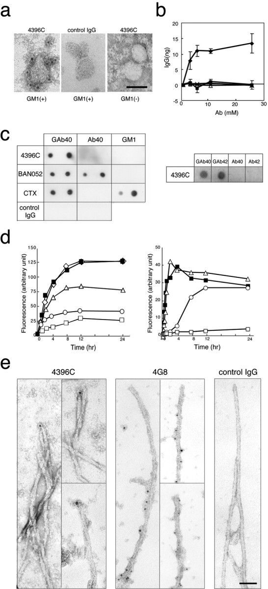
Characterization of the binding specificity of 4396C. a, Immunoelectron micrographs of liposomes. GM1-containing and GM1-lacking liposomes were subjected to immunoelectron microscopy of 4396C or isotype-matched control IgG staining after incubation with soluble Aβ40. GM1(+), GM1-containing liposomes; GM1(–), GM1-lacking liposomes. Scale bar, 50 nm. b, Quantitative assay of binding of 4396C to liposomes. GM1-containing liposomes were incubated with 4396C (diamonds) or isotype-matched control IgG (filled squares) after their mixing with soluble Aβ40 at indicated concentrations. GM1-lacking liposomes were also incubated with 4396C (triangles) or isotype-matched control IgG (× symbols). Ab, Antibody. c, Dot blot analysis. Left, Liposomes carrying GAβ (GAβ40), Aβ40, and GM1 in amounts equal to those contained in blotted liposomes (300 and 600 ng of Aβ40; 2 and 4 μg of GM1) were blotted. The blots were incubated with 4396C, BAN052, HRP-conjugated CTX, or isotype-matched control IgG. Right, Liposomes carrying GAβ (GAβ40 and GAβ42), prepared using Aβ40 or Aβ42, and Aβ40 and Aβ42 in amounts equal to those contained in GAβ40 and GAβ42 (600 ng of each peptide) were blotted. The blots were incubated with 4396C. d, Inhibition of amyloid fibril formation from soluble Aβ40 by 4396C. Left, Soluble Aβ40 was incubated with GM1-containing liposomes in the absence (filled squares) or presence of an antibody (4396C or 4G8). The molar ratios of 4396C to soluble Aβ40 were 0.3:50 (triangles), 1.3:50 (circles), and 4:50 (open squares) and that of 4G8 to Aβ40 was 4:50 (diamonds). Right, Soluble Aβ40 was incubated with preformed Aβ40 fibrils in the absence of an antibody (filled squares) or in the presence of 4396C. The molar ratios of 4396C to soluble Aβ40 were 0.3:50 (triangles), 1.3:50 (circles), and 4:50 (open squares). e, Immunoelectron micrographs of preformed Aβ40 fibrils. Aβ40 fibrils were formed by the extension reaction of Aβ40 seeds (10 μg/ml) with seed-free Aβ40 (50 μm), as described in Materials and Methods, and subjected to immunoelectron microscopy of 4396C, 4G8, or isotype-matched control IgG staining. Scale bar, 50 nm.