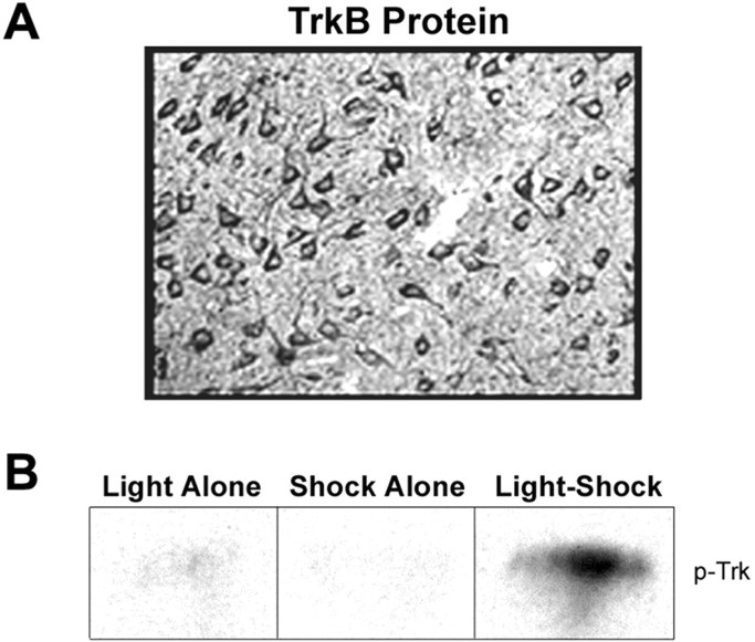
Figure 4.
Changes in levels of phosphorylated-Trk receptor protein in the amygdala after fear conditioning. A, Immunohistochemical analysis of TrkB immunoreactivity (high power) in the amygdala. B, Representative Western blots of amygdala samples probed with phosphorylated-Trk Ab. Samples were taken from animals that had been exposed to light presentations alone, shock-alone, or light-shock pairings.

