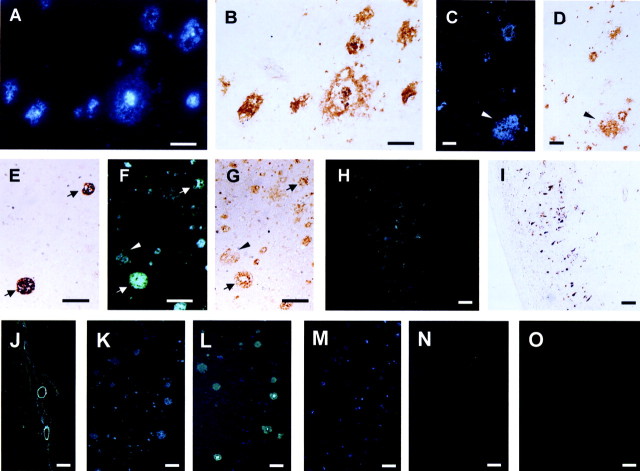
Figure 3.
Neuropathological staining of 6 μm AD brain sections from the temporal cortex (A-G, M, N) and hippocampus (H-L) and an aged normal temporal brain section (O). A-D are frozen sections, and E-O are paraffin-embedded sections. Many neuritic plaques are clearly stained with BF-168 (A). Intense fluorescence can be seen in the core of neuritic plaques. Aβ immunostaining with antibodies 6F/3D (B) in the adjacent section show an identical staining pattern of plaques. The brain section stained with BF-168 (C) and the adjacent section immunostained with a mAb against Aβ (D) demonstrate specific binding of BF-168 to diffuse amyloid plaques. Aβ1-40 (E) and Aβ1-42 (G) immunostaining and BF-168 staining (F) in serial sections reveal specific binding of BF-168 to not only Aβ1-40-positive plaques (arrows) but also Aβ1-42-positive diffuse plaques (arrowheads). H, BF-168-stained NFTs, and this staining correlated well with tau immunostaining (I) in the adjacent section. Clear staining of cerebrovascular amyloid (J) can also be observed. There is no obvious difference in the BF-168 staining ability in ethanol solution (K) and in the PBS solution (L) in the serial sections. BF-168 could not stain any plaques and tangles after the treatment with formic acid (N), in contrast to the staining without pretreatment with formic acid (M). Finally, no apparent staining was observed in aged normal brain sections (O). Scale bars: A-D, 50 μm; E-O, 100 μm.

