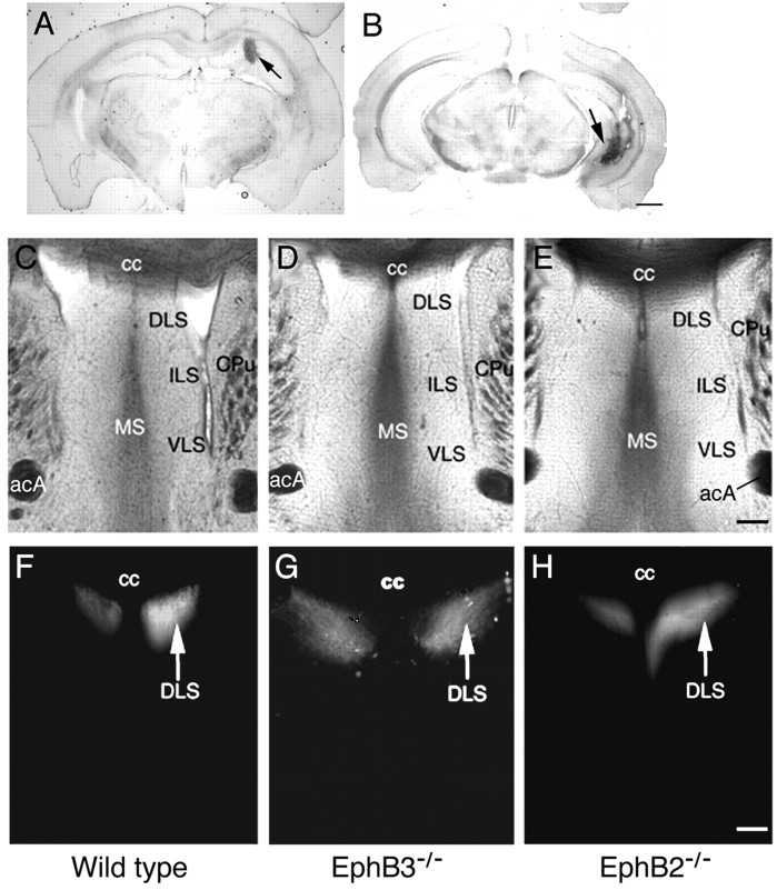
Figure 1.
Normal medial hippocampal projections to the lateral septum in EphB2- and EphB3-null mice. A, B, Typical Fluoro-ruby injection sites for hippocampal axon tracing in the medial hippocampus (A) and the lateral hippocampus (B). Injections were done according to stereotaxic coordinates by Franklin and Paxinos (1997). Arrows indicate injection sites. Scale bar, 1 mm. C-H, Tracing of the medial hippocampal axon terminal field in the lateral septum. C-E, Bright-field photomicrographs of sections of the traced brains containing the septal area from the wild-type (C), EphB3-/- (D), and EphB2-/- (E) mice. Major cytoarchitectural features are indicated. F-H, Dark-field photomicrographs depicting positions of the medial hippocampal axon terminals in wild-type (F), Eph B3-/- (G), and Eph B2-/- (H) mice. acA, Anterior commissure, anterior tract; cc, corpus callosum; CPu, caudate-putamen; DLS, dorsal lateral septum; ILS, intermediate lateral septum; MS, medial septum; VLS, ventral lateral septum. Scale bar, 0.2 mm.

