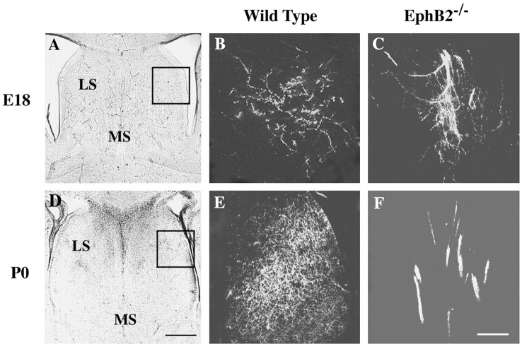
Figure 4.
Hippocampal axon defasciculation defects in embryonic and newborn EphB-null mice. DiI was placed in the lateral hippocampal regions. A, D, Bright-field photomicrographs of wild-type brain sections showing major cytoarchitectural features of the septal region. B, E, Defasciculated hippocampal axons in the lateral septum of wild-type mouse brains. C, F, Abnormal hippocampal axon bundles in the lateral septum of the EphB2-/- mutant mice. Images in B, C, E, and F are from the boxed area in A and D. Scale bars: B, 0.25 mm; F, 0.05 mm.

