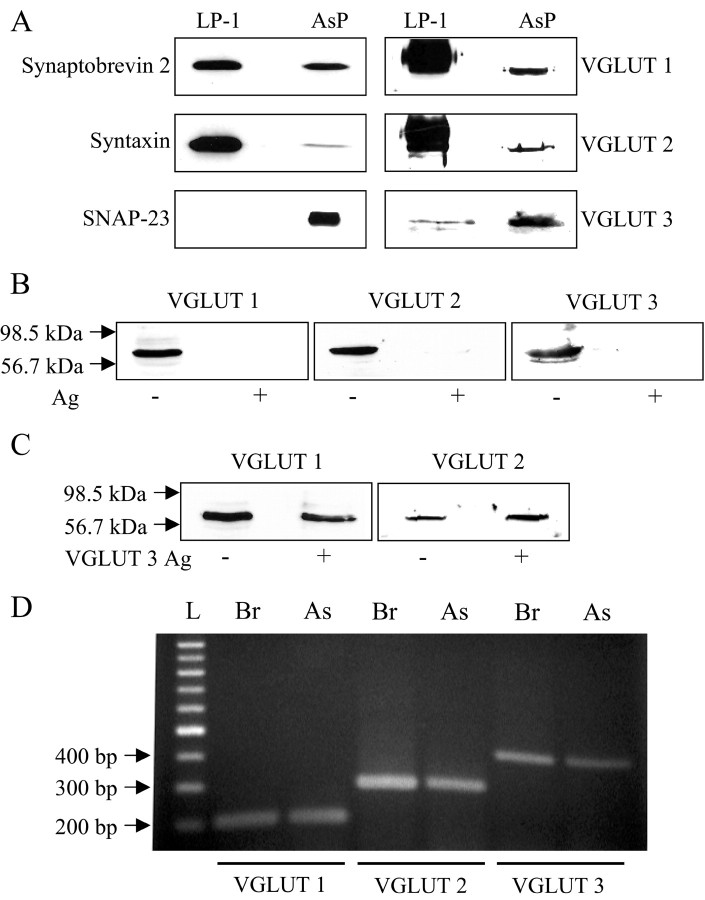
Figure 2.
Astrocytes in culture express VGLUTs 1 and 2. A, After SDS-PAGE, immunoblots indicate the presence of VGLUTs 1, 2, and 3 in astrocytes (AsP) as well as SNARE proteins, synaptobrevin 2, syntaxin, and SNAP-23. In LP-1 preparations, SNAP-23 was not detected. When probing SNARE proteins, LP-1 was loaded 1–2 μg per lane, whereas AsP was loaded 10–15 μg per lane. However, when probing VGLUTs, LP-1 and AsP were equally loaded at 15 μg per lane for VGLUTs 1 and 2 or 30μg per lane for VGLUT 3. B, The immunoreactive bands in AsP recognized by antibodies raised against VGLUT 1, 2, and 3 isoforms (Ag–) were completely abolished when antibodies were preincubated with their homologous antigens (Ag+). C, Antibodies raised against VGLUTs 1 and 2 do not cross-react with VGLUT 3 because their preadsorption with this heterologous antigen does not abolish the immunoreactive bands, although the same VGLUT 3 antigen in preadsorption with anti-VGLUT 3, as seen in B, abolishes its ability for detection. The arrows in B and C indicate molecular weight markers. D, RT-PCR using mRNA isolated from purified astrocytes (As) or from brain (Br) reveals the presence of VGLUT 1, 2, and 3 PCR products (207, 294, and 399 bp, respectively). L and arrows indicate molecular size markers (100 bp DNA ladder).

