
Figure 3.
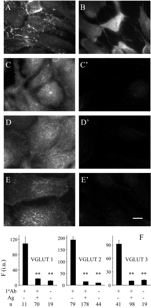
Subcellular localization of synaptobrevin 2 (A), SNAP-23 (B), VGLUT 1 (C), VGLUT 2 (D), and VGLUT 3 (E). VGLUT immunoreactivity was completely abolished when primary antibodies (1 oAb) were preadsorbed with their respective antigens (Ag) in cultured astrocytes (C′–E′, F). Scale bar: A, B, 20 μm; C–E, C′–E′, 10 μm. Fluorescent immunoreactivity is expressed in intensity units (i.u.). Bars represent means ± SEMs of measurements from the number of individual astrocytes (n). Asterisks indicate a significant change of measurements compared with the control group (1 oAb+, Ag–; one-way ANOVA, followed by post hoc Fisher's LSD test; **p < 0.01). We found no difference between measurements in preadsorption controls(1 oAb+, Ag+) when compared with controls in which primary antibodies were omitted (1 oAb–, Ag–).