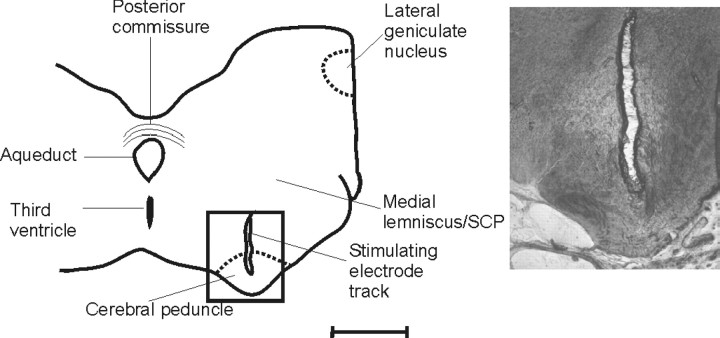
Figure 3.
Histological location of the CP stimulation site. Camera lucida drawing of a transverse section of the midbrain (BW5) at approximately AP +6.5. The inset shows a higher-power photomicrograph of a track made by the bipolar stimulating electrode. Scale bar, ∼3 mm for low-power outline; 1 mm for photomicrograph.

