
Figure 4.
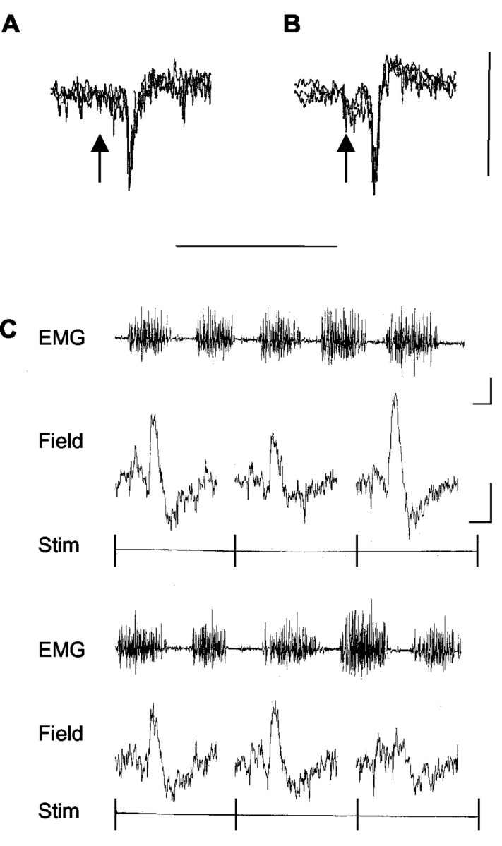
Sample climbing fiber field potentials. Extracellular recordings from the cerebellar cortex in the awake cat (3 sweeps superimposed) show sample climbing fiber field potentials obtained at the same paravermal recording site (in the molecular layer) and evoked by ipsilateral SR (A) or contralateral CP (B) stimulation. The stimulus was delivered at the time indicated by the arrow. Calibration: 0.2 mV, 50 msec. C, Evoked climbing fiber field potentials recorded during locomotion at a different paravermal recording site (located at or below the Purkinje cell layer). The EMG trace is continuous from the top left to the bottom right and displays activity in the ipsilateral forelimb extensor triceps brachii. Single-sweep traces below the EMG show individual cerebellar responses (field) evoked at 1.5 sec intervals by contralateral CP stimulation. Each single sweep is triggered at the onset of the stimulus (Stim). Calibration: EMG, 0.2 sec, 0.5 mV; field, 10 msec, 0.2 mV.