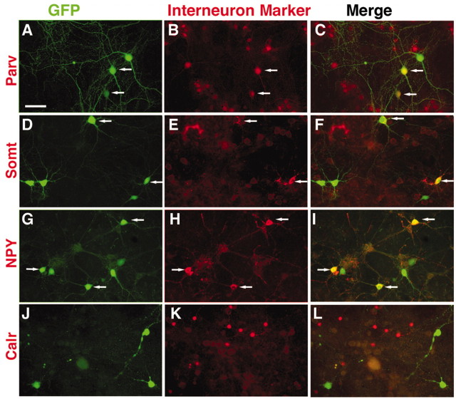
Figure 4.
Transplanted progenitors from the MGE give rise to distinct interneuron subtypes. In this experiment the E14.5 MGE donor cells were plated onto neonatal cortex feeders and cultured for 2 weeks (4 weeks in the case of parvalbumin). Each set of three panels shows identical fields, the left side of which shows GFP expression by the donor cells. The middle panels show expression of the interneuron subtype markers parvalbumin (Parv; B), somatostatin (Somt; E), neuropeptide Y (NPY; H), and calretinin (Calr; K). The panels on the right side show the merged images. Colabeling is present between some of the MGE donor cells and Parv, Somt, and NPY; however, MGE donors do not colabel for calretinin. Scale bar, 50 μm.

