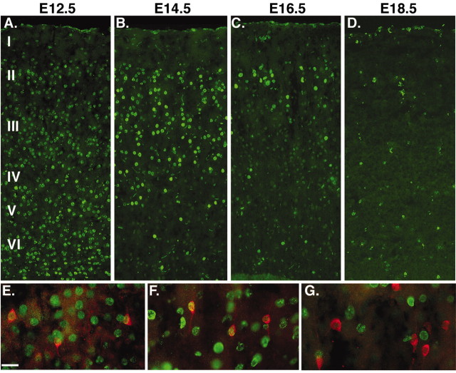
Figure 7.
Birth dating of calretinin-expressing cortical interneurons. A–D, Low-magnification views of BrdU staining in the somatosensory cortex of animals that received five injections of BrdU (50 mg/kg) 2 hr apart at E12.5 (A), E14.5 (B), E16.5 (D), or E18.5 (D). E, F, High-magnification views of calretinin (red) and BrdU (green) labeling in layer II from animals that received E12.5 (E), E14.5 (F), or E16.5 (G) injections. Most of the calretinin-expressing interneurons have a bipolar morphology similar to that seen in vitro and are labeled by BrdU injections at E12.5 (79%) or E14.5 (70%) but rarely later (only 1.8% at E16.5, and essentially none from injections at E18.5 or P1). Scale bar, 20 μm.

