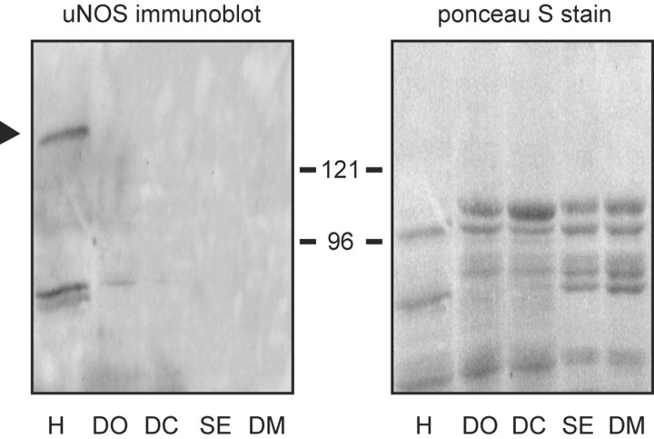
Figure 1.
Immunoblot analysis of NOS expression in lobster contractile tissues. Each lane contains 60 μg of cytosolic protein obtained, as indicated, from heart (H), walking leg dactyl opener muscle (DO), walking leg dactyl closer muscle (DC), superficial abdominal extensor muscle (SE), and dorsal thoracic body wall muscle (DM). To the left is an immunoblot generated with a uNOS antibody that recognizes a core domain conserved across a variety of vertebrate and invertebrate species. The black arrowhead denotes the position of the NOS-like protein. To the right is the Ponceau S stain of the same blot generated just before immunostaining, showing that equivalent protein loads were applied to each lane. Positions of molecular weight standards (in kilodaltons) are as indicated.

