
Figure 1.
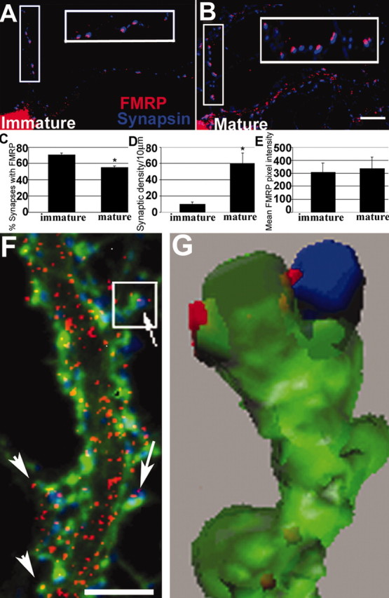
Developmental prevalence of FMRP at synapses. A, IF of an 8 DIV neuron shows frequent FMRP (red) colocalization with synapsin (blue; see enlarged inset). B, Same as A but a mature 19 DIV neuron. C, Histogram showing that 75% of synapsin puncta colocalized with FMRP at 8 DIV; only 54% colocalized at 19 DIV (*p < 0.05; Student's t test). D, Density of synapses increased as neurons matured (p < 0.05). E, There was no difference in FMRP levels in dendritic shafts of 8 and 19 DIV cultures. Scale bar, 10 μm. F, Triple-label IF detects FMRP (red), synapsin (blue), and F-actin (green). FMRP granules are distributed throughout dendrites, spine synapses (arrows), and filopodia (arrowheads). Scale bar, 5μm. G, 3-D reconstruction of boxed spine from F shows FMRP granules in spine neck and head, apposed to synapsin.