
Figure 5.
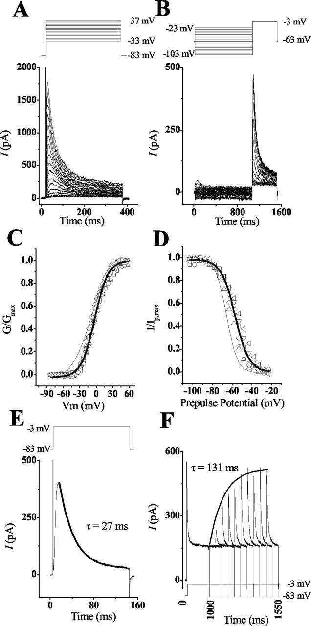
The activation and steady-state inactivation of cKv4.2 expressed in CHO cells differ from the kinetics in hair cells. A, C, Depolarization of transfected CHO cells elicited a transient voltage-dependent current that began to activate at -35 mV and was completely activated at 40 mV. To determine the conductance, the peak current amplitude was divided by the driving force across the membrane using the reversal potential of -83 mV (obtained from tail current measurements not shown). Conductance values were normalized to the maximal conductance and plotted against the membrane potential. The solid line was derived by fitting the data from these different cells to a Boltzmann equation (Fig. 3). From this fit, the midpoint (V1/2) and slope (k) parameters were derived as -2.8 ± 0.7 and 12.3 ± 0.6 mV, respectively. For comparison of CHO versus hair cell responses, the voltage-conductance fit from Figure 3B (thin line) is superimposed on this plot. B, D, Steady-state inactivation properties of the transient current were determined by presenting a prepulse of 1 sec, at the given membrane potentials, followed by a test pulse of -3 mV for 450 msec. The peak current during the test pulse was normalized to the maximal current obtained after a prepulse of -103 mV. The solid line was derived using a Boltzmann equation (Fig. 3). This fit generated midpoint (V1/2) and slope (k) values of -57.95 ± 0.55 and -7.6 ± 0.5 mV, respectively. For comparison of CHO versus hair cell responses, the steady-state inactivation fit from Figure 3F (thin line) is superimposed on this plot. E, Plot of the response that was generated by depolarizing a CHO cell to -3 mV from a holding potential of -83 mV. When these responses were fit with a single-order exponential, the rate of current inactivation (τ = 27 msec) did not differ significantly from that seen in hair cells. F, The recovery from inactivation for recombinant channels was longer than recovery for native channels. CHO cells were voltage clamped to -83 mV and depolarized to -3 mV for 1 sec to fully inactivate transient currents. A prepulse to -83 mV of increasing duration (50 msec intervals) was given to deinactivate the current, followed by a depolarizing test pulse to -3 mV to activate the deinactivated transient channels. The rate of recovery from inactivation was determined by plotting the peak IA generated against the prepulse duration. The solid line shows the curve fit generated for the responses using the equation described in Figure 4. This fit generated the exponential time constant τ = 131 msec.