
Figure 7.
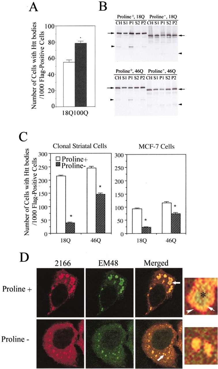
Effects of polyglutamine and polyproline regions on the formation of htt bodies in MCF-7 cells and clonal striatal cells (x57). A, The number of MCF-7 cells with htt bodies per 1000 Flag-positive cells is significantly increased in cultures transfected with FH969-100 compared with cultures transfected with FH969-18. One thousand Flag-positive cells were analyzed on each slide. *p < 0.05 (Student's t test; n = 6 slides). B, Western blots of htt expression in subcellular fractions prepared from cells transfected with FH969-18 and FH969-46 with (+) and without (-) the encoded polyproline region. Levels of intact htt (arrows) are approximately the same after polyproline deletion in the crude homogenate (CH), soluble (S1, S2) and membrane (P2) fractions. The P1 fraction shows a small decline in htt levels after proline deletion. Proline deletion changes the size of Flag-htt based on its faster migration on SDS-PAGE. N-htt fragments (arrowhead) still form after polyproline deletion, although proline removal in wt htt alters the pattern of breakdown products. C, x57 and MCF-7 cells were transfected with FH969-18 or FH969-46 with or without the polyproline region in htt, and the formation of htt bodies was analyzed 24 hr after transfection as in A. Note that a greater proportion of clonal striatal cells form htt bodies than MCF-7 cells. There is a significant reduction in the number of cells that form htt bodies when the polyproline region is removed (proline-) from wt or mutant htt in both cells lines (Student's t test; *p < 0.05). D, Immunofluorescence labeling for htt bodies in MCF-7 cells with antibodies 2166 and EM48 reveals changes in the morphological appearance of htt-labeled dense structures when the polyproline region is removed (proline-). With the intact polyproline region (proline+) in htt, htt epitopes in htt bodies segregate into a shell (red, arrowhead) and core (green, asterisk) and overlap in an intermediate region (yellow, arrow) as shown in the enlarged htt body from the merged image and also in Figure 1C. With polyproline deletion (proline-), more diffuse htt occurs in the cytoplasm, and htt epitopes do not partition into shell and core domains but codistribute in patches (see enlargement from merged image). Some patches are smaller than htt bodies.