Figure 1.

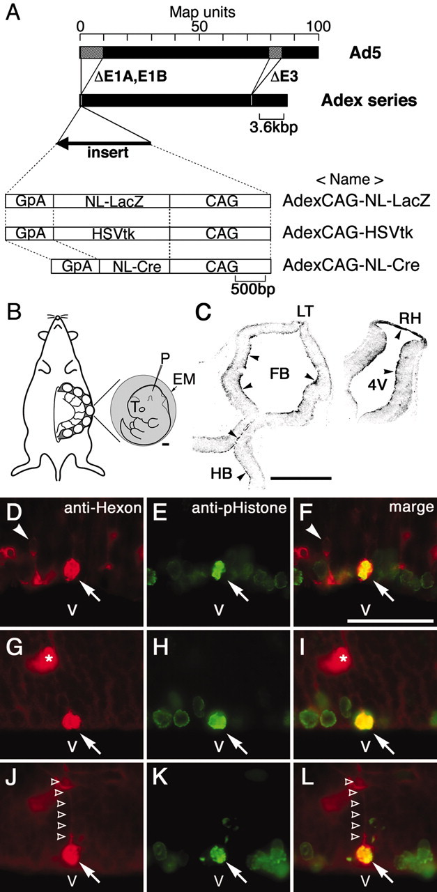
Construction of adenoviral vectors and adenoviral injection. A, Replication-incompetent adenoviral vectors (Adex series) that lack the E1A, E1B, and E3 region were generated from human Ad5. An expression unit was inserted into the E1A-E1B deleted region. AdexCAG-NL-LacZ expresses NL-LacZ. AdexCAG-HSVtk expresses HSVtk as a suicide gene. AdexCAG-NL-Cre expresses NL-Cre. B, Embryos were manipulated by exo utero surgery (see Materials and Methods). Exposed embryos (E10.5) lapped with external embryonic membrane (EM: yolk sac and amnion) are illustrated. Scale bar, 500 μm. Adenoviral vectors were injected into the midbrain ventricle with a heat-pulled glass pipette (P). C, Five hours after injection of AdexCAG-NL-LacZ into E10.5 embryo, brains were sectioned transversely and stained with polyclonal anti-hexon antibody. Anti-hexon immunoreactivity was observed along the ventricular surfaces (arrowheads). Scale bar, 500 μm. D-L, Two hours after injection of AdexCAG-NL-LacZ into E11.5 embryos, brains were sectioned transversely and double-immunostained with monoclonal anti-hexon (indicated in red; D, G, J) and, as a mitotic-cell marker, polyclonal anti-pHistone (indicated in green; E, H, K). F, I, and L indicate the merged view of D and E, G and H, and J and K, respectively. Scale bar, 50 μm. Arrows indicate the cell bodies of hexon- and pHistone-positive cells. The hexon- and pHistone-positive cells in D-F and G-L may be undergoing metaphase and anaphase, respectively. The hexon-and pHistone-positive cell in J-L has a short ascending process (unfilled arrowheads). Arrowheads in D and F indicate a hexon-positive cell in G1 phase that appears to be migrating away from the ventricular surface. Asterisks indicate blood vessels. 4V, Fourth ventricle; CAG, ubiquitous and strong promoter; GpA, poly-A region of rabbit β-globin; T, telencephalon; FB, forebrain ventricle; HB, hindbrain ventricle; LT, lamina terminalis; RH, roof of hindbrain; V, ventricle.
