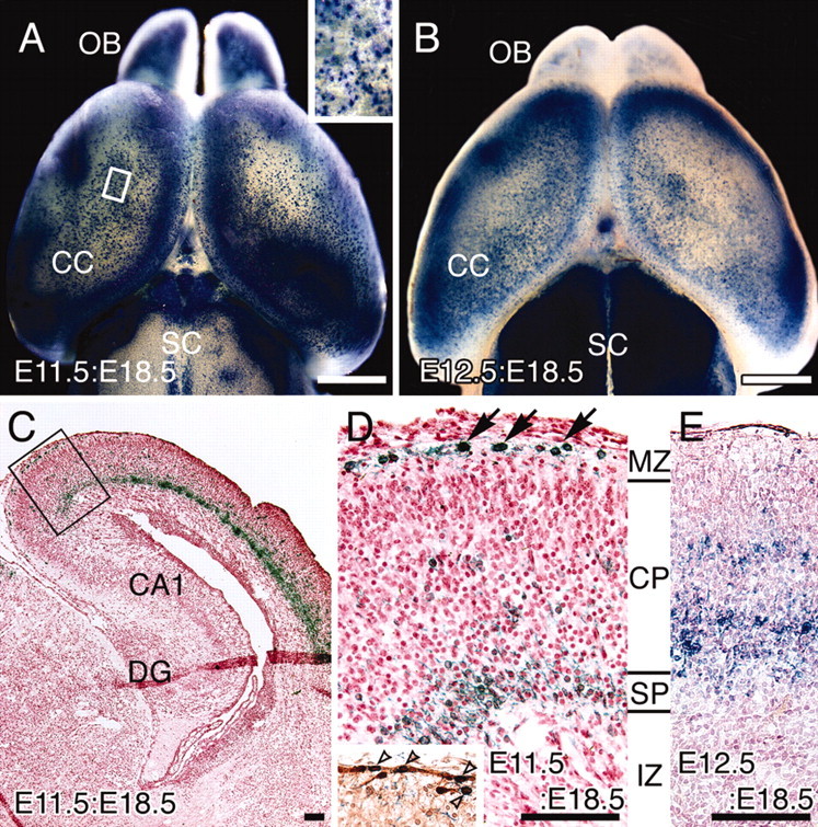
Figure 2.

Distribution of β-gal-positive cells in E11.5:E18.5 and E12.5:E18.5 brains. AdexCAG-NL-LacZ was injected into the midbrain ventricles of embryos at E11.5 and E12.5. At E18.5, each manipulated brain was stained for β-gal by whole mount. Dorsal views of the E11.5:E18.5 (A) and E12.5:E18.5 (B) brains are indicated. A high-magnification view of the area indicated by the white box in A is shown as an inset at the top right of A. Transverse cryosections from E11.5:E18.5 (C, D) and E12.5:E18.5 (E) brains were counterstained with neutral red. A high-magnification view of the box in C is shown in D. The β-gal-positive cells in E11.5:E18.5 brains are located in the marginal zone (MZ; arrows in D) and subplate (SP). The inset in D indicates a neighboring section of D stained with anti-calretinin antibody. Unfilled arrowheads indicate β-gal- and calretinin-positive cells in the marginal zone of a E11.5:E18.5 brain. β-gal-positive cells in E12.5:E18.5 brains (E) are located in the CP. CA1, Hippocampal CA1 region; DG, dentate gyrus; IZ, intermediate zone; OB, olfactory bulb; SC, superior coliculus. Scale bars: A, B, 1 mm; C-E, 100 μm.
