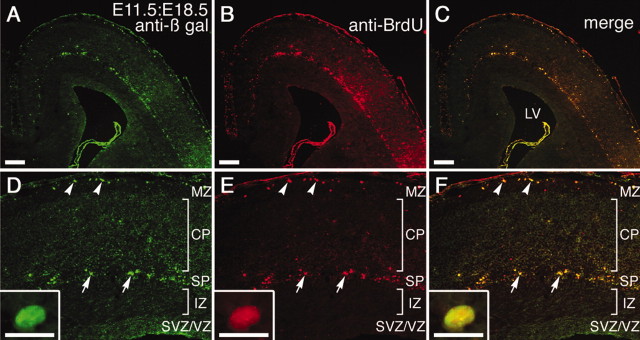
Figure 3.
BrdU labeling of an E11.5:E18.5 brain. A mixture of AdexCAG-NL-LacZ and BrdU was injected into the midbrain ventricle of an embryo on E11.5. At E18.5, the E11.5:E18.5 brain was sectioned transversely and double-immunostained with anti-β-gal (shown in green; A, D) and anti-BrdU (shown in red; B, E) antibodies. Arrowheads and arrows indicate double-positive C-R cells and subplate (SP) neurons, respectively. A high-magnification view of the double-positive subplate neuron is shown in insets in D-F. The CP and intermediate zone (IZ) are almost entirely β-gal- and BrdU-negative. LV, Lateral ventricle; MZ, marginal zone. Scale bars: 200 μm; insets, 20 μm.

