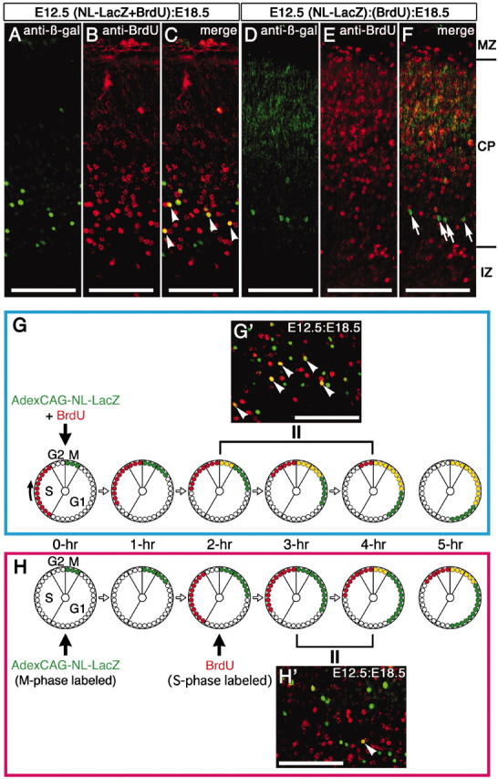
Figure 4.

Feature of adenovirus-mediated gene transfer into embryonic brain. AdexCAG-NL-LacZ and BrdU were simultaneously injected into the midbrain ventricle of E12.5 mouse embryos (A-C, G′). In the other cases, AdexCAG-NL-LacZ and BrdU were injected at separate times (D-F, H′): first, AdexCAG-NL-LacZ was injected into the midbrain ventricle of E12.5 embryos, and then 15 (D-F) or 2 (H′) hr after the adenoviral injection, BrdU was injected into the abdominal cavity of the manipulated pregnant dams. At E18.5, the embryos were fixed and sectioned transversely on a cryostat. The transverse sections were double-immunostained with anti-β-gal and anti-BrdU antibodies. After the simultaneous injection, the following cell-labeling patterns were seen in the cortical plate of the E12.5:E18.5 brain (C, G′): β-gal-positive cells (shown in green), BrdU-positive cells (shown in red), and β-gal- and BrdU-double-positive cells (shown in yellow). In contrast, after injection at separate times, the population of double-positive cells (shown in yellow) in the E12.5:E18.5 brains (F, H′) was not observed (F) or was greatly reduced (H′). The diagram in G indicates the labeling schedule for the experiment using simultaneous injection of BrdU and adenoviral vectors. Arrowheads in C, G′, and H′ indicate β-gal- and BrdU-double positive cells (shown in yellow). Arrows in F indicate β-gal-single-positive cells. The diagram in H indicates the labeling schedule for the experiment in which BrdU and adenoviral vector were applied at different times. One cell cycle is illustrated by a large circle, as shown at the lower left in G. The cell cycle progresses in a single direction (from G1 to S, G2, and M phases). The small red-filled circles indicate the fractions of proliferating cells that are labeled with BrdU during S-phase. The small green-filled circles indicate the fractions of proliferating cells that are infected with AdexCAG-NL-LacZ during M-phase and were found with labeling to be positive for β-gal. The small yellow-filled circles indicate the BrdU- and β-gal-double-positive cells. One small circle is equivalent to ∼20 min on one cell cycle. IZ, Intermediate zone; MZ, marginal zone. Scale bar, 100 μm.
