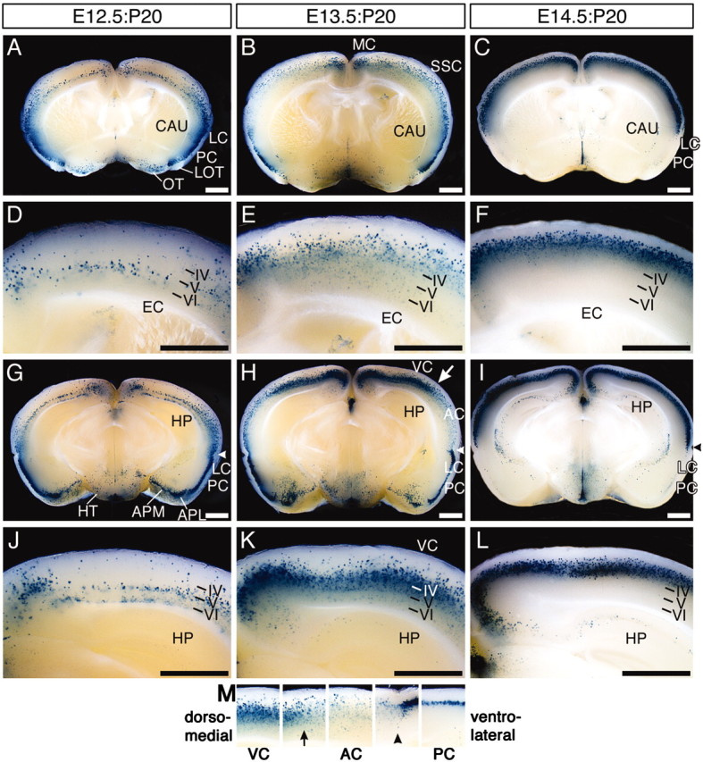
Figure 5.

Transverse sections of E12.5:P20, E13.5:P20, and E14.5:P20 brains. E12.5:P20 (left column), E13.5:P20 (middle column), and E14.5:P20 (right column) brains were sectioned transversely. A-F and G-L indicate the anterior and posterior regions of each brain, respectively. Arrowheads inG-I and M indicate the rhinal fissure. The boundary between the dorsal part of the auditory cortex and the lateral part of the secondary visual cortex in the E13.5:P20 brain is indicated by the white arrow in H and the arrow in M. Each cortical region in H is arranged in line in M. IV-VI, The layer number in the cerebral cortex; AC, primary auditory cortex; EC, external capsule; HP, hippocampus; HT, hypothalamus; LOT, lateral olfactory tract; MC, primary motor cortex; OT, olfactory tubercle; SSC, primary somatosensory cortex; VC, primary visual cortex. Scale bar, 1 mm.
