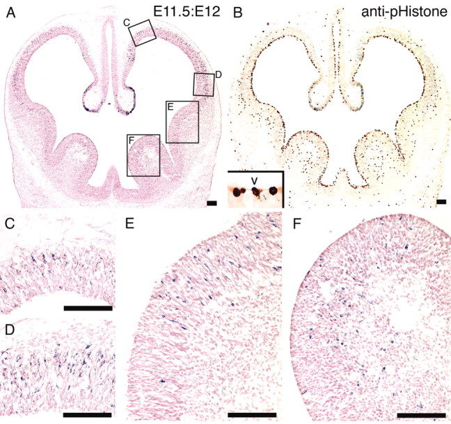
Figure 9.

Distribution of β-gal-positive cells in E11.5:E12 brain. AdexCAG-NL-LacZ was injected into the midbrain ventricle of the E11.5 embryos. Twelve hours (E12) after the adenoviral injection, the embryos were fixed and sectioned transversely. The sections were stained for β-gal and with neutral red (A). C and D are high-magnification views of the dorsomedial and ventrolateral regions of the cerebral cortex sections shown in A. E and F are high-magnification views of the medial and lateral ganglionic eminences from A. The neighboring sections were stained for β-gal and, as a mitotic cell marker, with anti-pHistone antibody (B). The nuclei and cell bodies of progenitor cells undergoing mitosis are located on the ventricular surface, and their cell bodies are swollen and exposed to the ventricle (B, inset). V, Ventricle. Scale bar, 100 μm.
