
Figure 12.
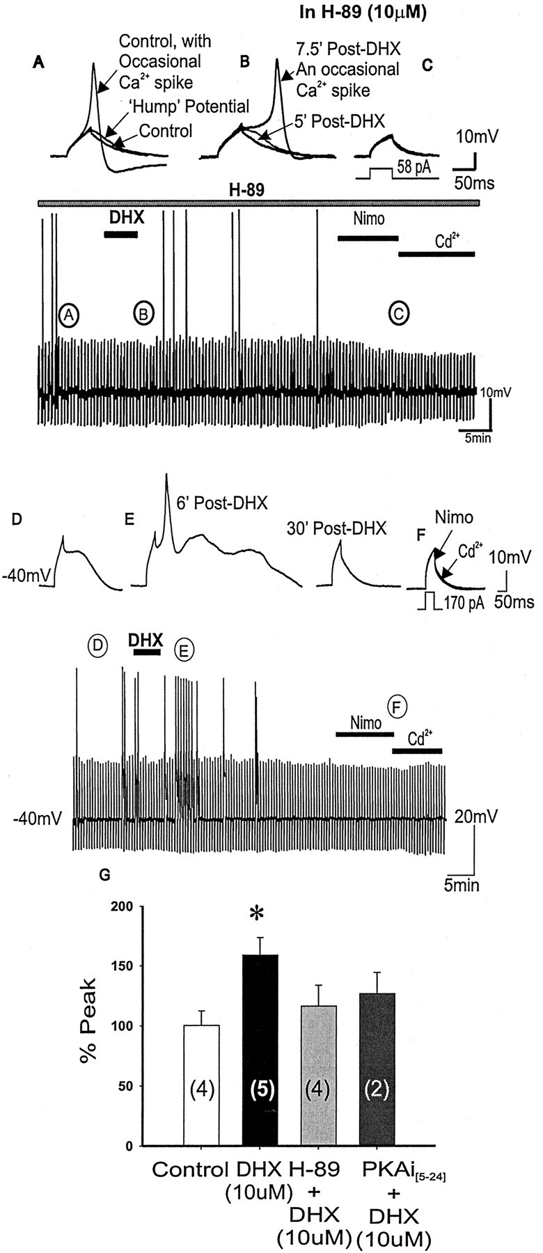
Blocking PKA activation by H-89 or PKAi[5-24] prevented D1/D5R-mediated enhancement of the evoked subthreshold Ca2+ hump potential. A-C, Representative traces corresponding to the continuous time-compressed traces of evoked subthreshold Ca2+ potentials or occasional evoked Ca2+ spikes with application of the specific PKA inhibitor H-89 alone. Subthreshold Ca2+ potentials or occasional Ca2+ spikes are observed in the control state (A), suggesting that spike firing properties are unchanged with H-89 (10 μm) application. After DHX application (B), the frequency of Ca2+ spikes is unchanged and no potentiation is observed, suggesting that D1/D5R potentiation of subthreshold Ca2+ hump potentials depends on PKA activation. Further addition of nimodipine and cadmium (C) resulted in membrane capacitive responses. Bottom time-compressed traces indicate the time course of the response. D-F, When using PKAi[5-24] (10 μm) in the patch pipette, application of DHX resulted in blocking most of the expected potentiation by the D1 agonist (E); however, a few hump potentials could still be potentiated to Ca2+ spikes (E). Further addition of nimodipine and cadmium (F) resulted in membrane capacitive responses. G, Histograms summarizing group data showing that although D1/D5R activation (with DHX alone) potentiated subthreshold Ca2+ plateaus, D1/D5R activation in the presence of PKA inhibitor H-89 or PKAi[5-24] (10 μm) in the patch pipette did not potentiate the Ca2+ potentials, suggesting that D1/D5R-mediated potentiation depends on PKA activation. The bottom time-compressed traces indicate the time course of the response.