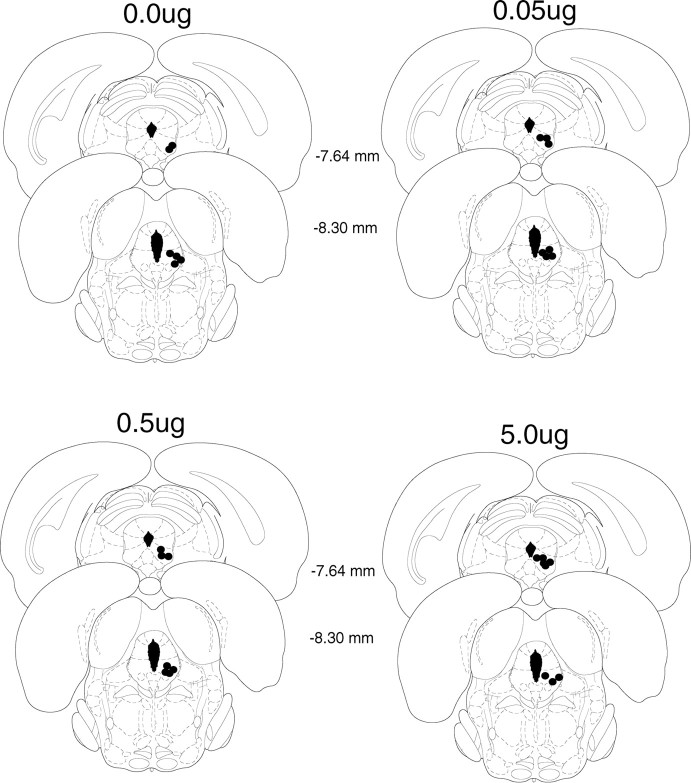
Figure 4.
Cannula placement in the periaqueductal gray. Illustration of injection cannula placements in the periaqueductal gray for experiment 4 is shown. Placements represented are from all rats included in the final analysis. Atlas templates were adapted from Paxinos and Watson (1998) (distances in millimeters from bregma).

