
Figure 6.
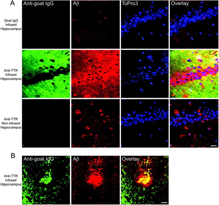
Chronic infusion of an anti-TTR antibody into the hippocampus of APPSw mice results in antibody deposition and Aβ accumulation within the infused hippocampus. Laser scanning confocal analysis was performed on hippocampal sections double immunolabeled for goat IgG or TTR (anti-goat IgG; green) and Aβ (4G8; red). Yellow indicates regions of costaining. The DNA-binding dye ToPro3 (blue) was used to stain nuclei. A, An anti-goat IgG antibody reveals that control goat IgG infused into the hippocampus is cleared after the 2 week infusion. Some Aβ staining occurs within the extracellular space and around the CA1 neurons in goat IgG-infused hippocampi and in the noninfused hippocampi of the anti-TTR-infused mice. However, mice infused with the anti-TTR antibody demonstrated a dramatic deposition of the anti-TTR antibody within the extracellular space of the infused, but not the noninfused, hippocampi. In addition, infusion of the anti-TTR antibody increased the amount of Aβ around and within the CA1 neurons near the infusion site. Scale bar, 20 μm. B, The anti-TTR antibody colabeled Aβ plaques in the infused hippocampus. Scale bar, 20 μm.