Figure 7.

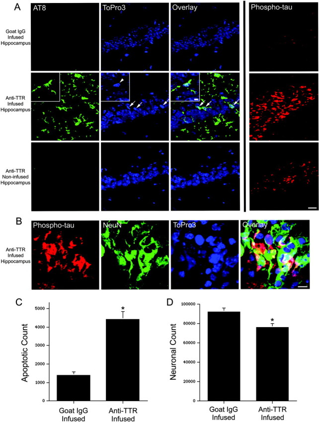
Chronic infusion of an anti-TTR antibody into the hippocampus of APPSw mice results in tau phosphorylation, apoptosis, and neuronal loss within the infused hippocampus. A, After a 2 week infusion of goat IgG, no phosphorylated tau [AT8, green; or phospho-tau(Thr231), red] is observed within the hippocampal neuronal fields. The CA1 region is shown, and the DNA-binding dye ToPro3 (blue) was used to stain nuclei. In contrast, a 2 week infusion of an antibody to TTR resulted in AT8 staining within occasional CA1 neurons. Many of these neurons possess pyknotic nuclei (ToPro3, blue; arrows). The inset shows a higher magnification within the CA1 neuronal field of a phosphorylated tau-stained neuron with a condensed nucleus (arrowhead) and beaded processes indicative of degeneration. Many neurons within the CA1 field also stain intensely for tau phosphorylated at Thr231 (phospho-tau, red). No AT8 staining and little phospho-tau(Thr231) is observed in the noninfused hippocampus. Scale bar, 20 μm; inset, 5 μm. B, CA1 neurons within the anti-TTR-infused hippocampus stain intensely for phospho-tau(Thr231) (red) and the neuronal marker NeuN (green). Scale bar, 10 μm. C, Unbiased stereology was used to determine the total number of cells with pyknotic nuclei within the infused CA1 pyramidal neuronal fields of goat IgG- (n = 4) and anti-TTR- (n = 4) infused mice. *p < 0.01, two-tailed Wilcoxon signed rank test. D, The total number of CA1 neurons was significantly reduced in anti-TTR-(n = 4) compared with goat IgG-(n = 4) infused mice. *p < 0.05, two-tailed Wilcoxon signed rank test.
