
Figure 1.
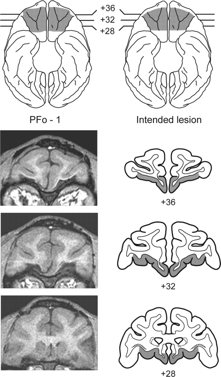
Top, Ventral view of a standard rhesus monkey brain showing the location and extent of the PFo lesion for monkey PFo-1 (left; shaded region) and the extent of the intended PFo lesion (right; shaded region). Bottom, Representative T1-weighted MR images from PFo-1 (left) and coronal sections at matching levels from a standard rhesus monkey brain (right) showing the extent of the intended removal. The numerals indicate the distance in millimeters from the interaural plane (0).