
Figure 6.
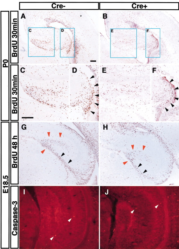
Decreased proliferation but normal survival in the Fgfr1Δflox hippocampus. A-F, Proliferating cells identified by a 2 hr BrdU incorporation at P0. C-F show a 2× higher magnification of blue squares in A and B. Proliferating cells in DG (C, E) and in hippocampal VZ (D, F, black arrowheads) are greatly decreased in hGFAP-Cre/+; Fgfr1flox/flox mutants. G, H, BrdU was injected at E16.5, and embryos were collected at E18.5. The number of cells along the migrating pathway to the DG (black arrowheads) as well as cells that are locally proliferating in the DG (red arrowheads) are clearly decreased in hGFAP-Cre/+; Fgfr1flox/flox mutants. I, J, Apoptotic cells (white arrowheads) are detected by an antibody for cleaved Caspase-3, revealing no major differences between Cre- and Cre+ mice at E18.5. Cre-, Fgfr1flox/flox; Cre+, hGFAP-Cre/+; Fgfr1flox/flox. Scale bar, 200 μm.