Figure 4.
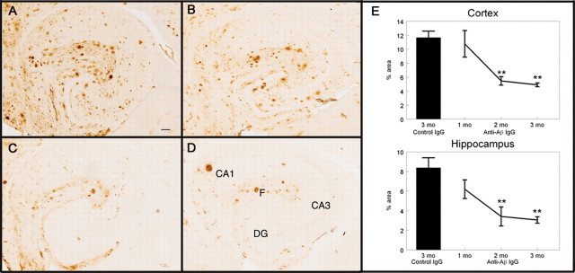
Total Aβ immunohistochemistry is reduced after 2 months of systemic anti-Aβ antibody administration. A-D, Total Aβ immunohistochemistry in the hippocampus of APP transgenic mice receiving the control antibody for 3 months (A; percent area for this section was 9.12%), the anti-Aβ antibody for 1 month (B; percent area for this section was 6.84%), the anti-Aβ antibody for 2 months (C; percent area for this section was 3.23%), or the anti-Aβ antibody for 3 months (D; percent area for this section was 2.49%). Magnification, 40×. Scale bar, 120 μm. E, Quantification of the percent area occupied by the Aβ-positive stain in the frontal cortex and hippocampus. The single bar shows the value for APP transgenic mice receiving the control antibody for 3 months. The line shows the values for APP transgenic mice receiving the anti-Aβ antibody for 1, 2, and 3 months. **p < 0.01. CA1, Cornu ammonis 1; CA3, cornu ammonis 3; DG, dentate gyrus; F, hippocampal figure.

