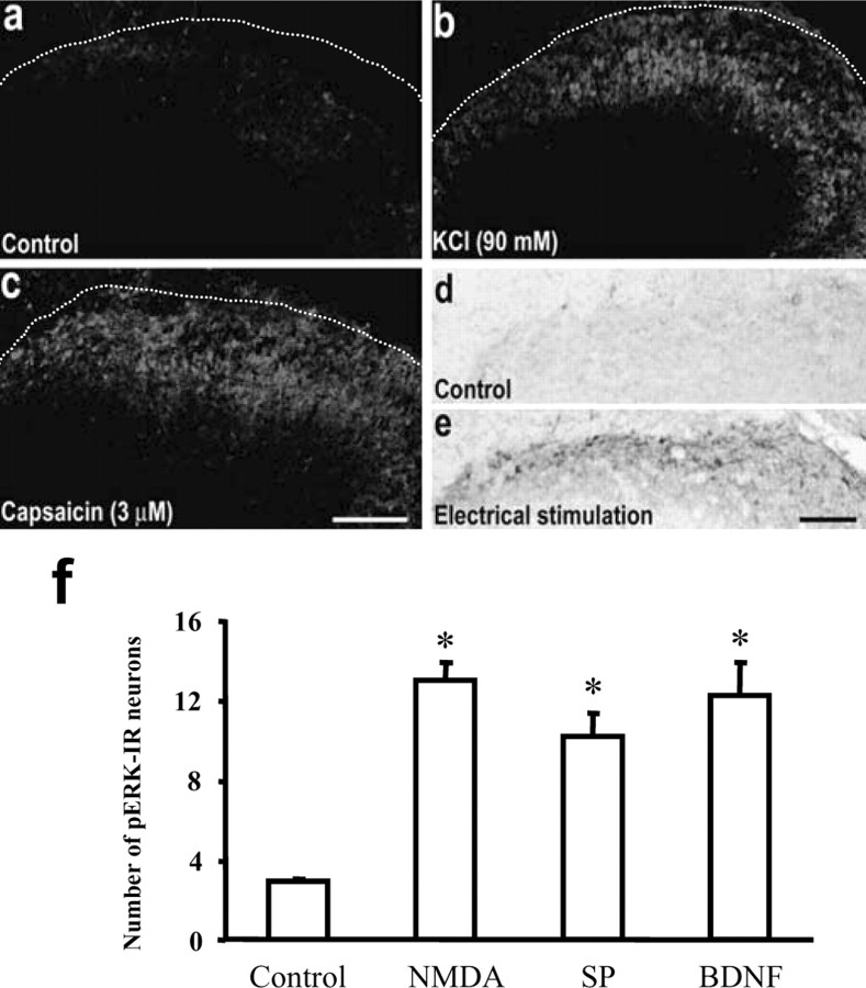
Figure 2.
a–c, pERK immunofluorescence in control unstimulated (a), KCl depolarized (b), and capsaicin-stimulated (c) spinal cord slices. The slices were fixed 10 min after the capsaicin exposure (3μm; 5 min) and KCl (90 mm; 5 min) depolarization. The dotted lines indicate the top border of the gray matter of the spinal cord. d, e, pERK immunostaining by DAB in control unstimulated and electrical C-fiber stimulation (1 mA, 50 Hz; 50 msec; 150 pulses) of an attached dorsal root in spinal cord slices. The slices were fixed 2 min after electrical stimulation. Scale bars, 100μm. f, Numbers of pERK-IR neurons after exposure of spinal cord slices to NMDA (100 μm), substance P (SP; 100 μm), and BDNF (200 ng/ml). ANOVA indicates an overall effect of these agents on pERK expression (F(3, 11) = 18.489; p < 0.0001). *p < 0.01, compared with control (n = 4–5). All of the slices were fixed 10 min after exposure.

