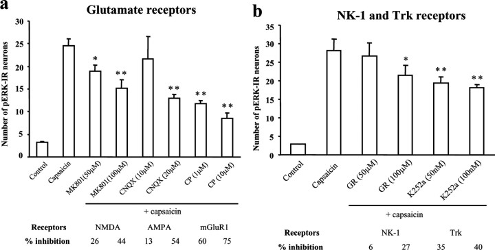
Figure 4.
a, b, Involvement of ionotropic, metabotropic, and tyrosine kinase receptors in capsaicin-induced pERK induction in spinal cord slices. a, Number of pERK-IR neurons in the superficial dorsal horn (laminas I-II) after capsaicin stimulation (3 μm; 5 min) in the presence of the NMDA receptor antagonist MK-801 (50 and 100 μm), the AMPA receptor antagonist CNQX (10 and 20μm), and mGluR1 antagonist CPCCOEt (CP; 1 and 10μm). ANOVA indicates an overall effect of these agents on pERK expression (F(7, 33) = 29.782; p < 0.0001). Mean ± SEM; *p < 0.05; **p < 0.01, compared with capsaicin (n = 4–6). b, Number of capsaicin-induced pERK-IR neurons in the presence of the NK-1 receptor antagonist GR205171A (GR; 50 and 100 μm) and the Trk inhibitor K252a (50 and 100 nm). ANOVA indicates an overall effect of these agents on pERK expression (F(5, 25) = 16.442; p < 0.0001). *p < 0.05; **p < 0.01, compared with capsaicin (n = 4–6). Six to eight sections (15μm) were included for each slice. The slices were incubated with each drug for 5–10 min and fixed 10 min after the capsaicin stimulation.

