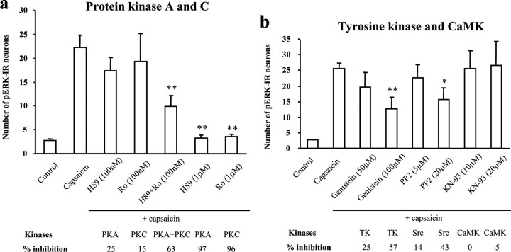
Figure 6.
a, b, Involvement of PKA, PKC, Src, and CaMK in capsaicin-induced pERK induction in spinal cord slices. a, Numbers of pERK-IR neurons in the superficial dorsal horn (laminas I-II) after capsaicin stimulation (3 μm; 5 min) in the presence of the PKA inhibitor H89 (0.1 and 1 μm), the PKC inhibitor Ro-31–84-25 (Ro; 0.1 and 1 μm), and H89 plus Ro-31–84-25 (0.1 μm). ANOVA indicates an overall effect of these inhibitors on pERK expression (F(6, 27) = 14.438; p < 0.0001). **p < 0.01, compared with capsaicin. Mean ± SEM (n = 3–6). b, Numbers of capsaicin-induced pERK-IR neurons in the presence of the general tyrosine kinase(TK) inhibitor genistein (50 and 100μm), the Srcinhibitor PP2 (5 and 20μm), and the CaMK inhibitor KN93 (20 μm).ANOVA indicates an overall effect of these agents on pERK expression (F(7, 23) = 4.327; p = 0.003). *p < 0.05; **p < 0.01, compared with capsaicin (n=3–6).Six to eight sections (15 μm) were analyzed for each slice. Mean ± SEM. The slices were incubated with each drug for 10 min and fixed 10 min after the capsaicin stimulation.

