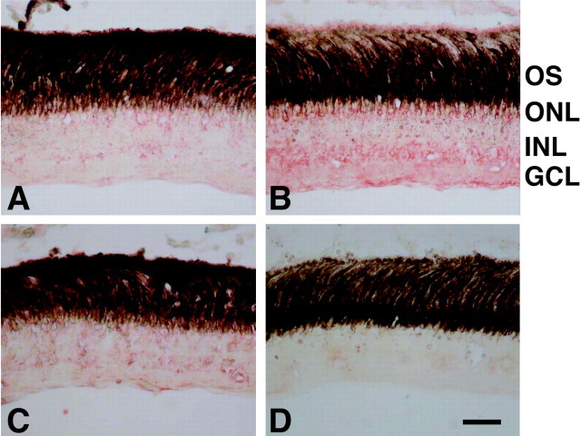
Figure 5.
Immunohistochemical study of purpurin in the retina. A–D, Retinal sections from various days after optic nerve transection were incubated with the anti-peptide antibody for purpurin (A–C) and preimmune serum (D). The red immunoreactive signals of purpurin increased in all of the nuclear layers, including the ganglion cell layer (GCL) 5 d (B) after optic nerve section compared with the control (A). Immunoreactivity decreased by 20 d after optic nerve transection (C). No immunoreactivity could be seen in the retina 5 d after optic nerve transection with preimmune serum (D). INL, Inner nuclear layer; GCL, ganglion cell layer; OS, outer segment. Scale bar, 20 μm.

