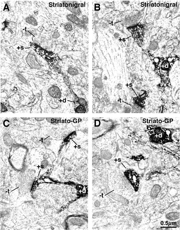
Figure 4.

Electron microscopic images of dendrite (+d) and spine (+s) labeling of striatonigral (A, B) and striato-GP (C, D) neurons that had been retrogradely labeled with BDA3k. Note that the labeled striatonigral (A, B) spines receive asymmetric synaptic contact from smaller unlabeled terminals (-t) than do the striato-GP neuron spines (C, D).
