
Figure 5.
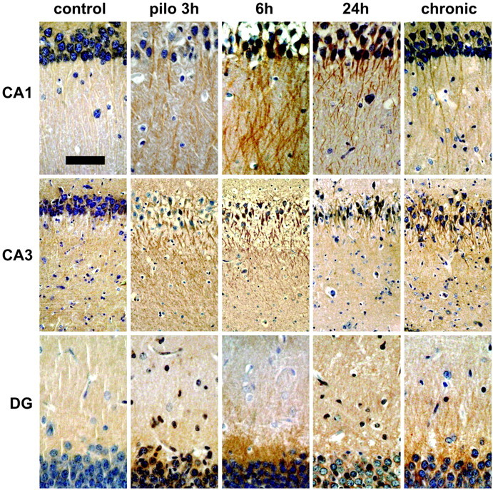
Pilocarpine seizure-induced dendritic BDNF mRNA is translated into protein. Shown are representative hematoxylin counterstained coronal brain sections at the level of the dorsal hippocampus (plate 39) (Pellegrino et al., 1979), exhibiting DAB-labeled BDNF-like immunoreactivity (LI). Omitting the primary antibody to estimate nonspecific signal yielded completely negative labeling (data not shown). Under control conditions (control), BDNF-LI is localized in the proximal dendrites. Peaking 6 hr after pilocarpine injection (pilo 6h; 300 mg/kg, i.p.), BDNF-LI is found in the stratum radiatum of CA1 pyramidal neurons, in the stratum lucidum, and in the radiatum of CA3 pyramidal neurons and in the proximal third of dentate gyrus granule cell dendrites. Increased dendritic BDNF-LI is also observed in spontaneously seizing animals killed 3-4 weeks after pilocapine administration. Scale bar: CA1 and DG, 75 μm; CA3, 120 μm.