
Figure 6.
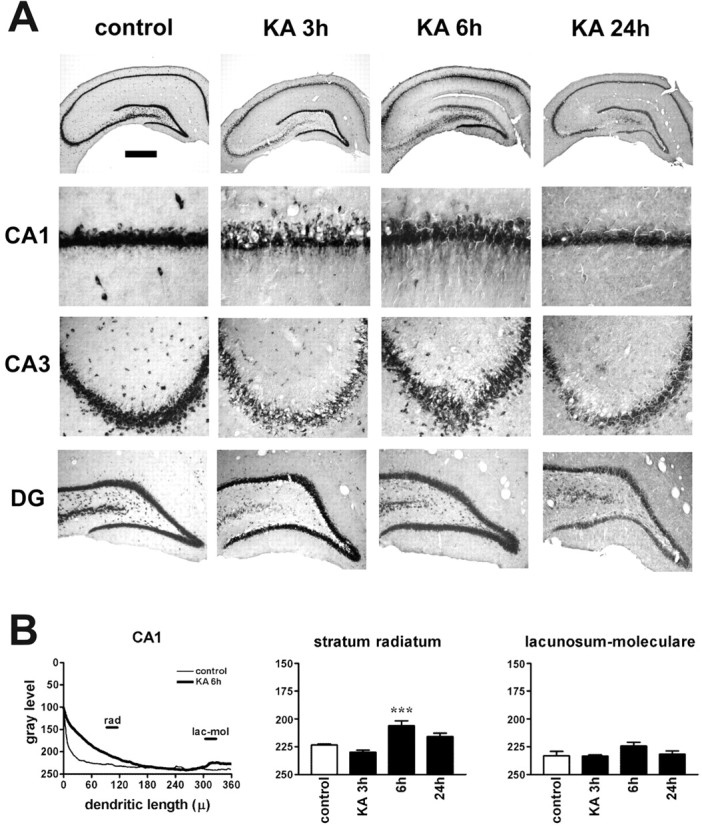
Kainate seizures induce dendritic targeting of BDNF mRNA in CA1 pyramidal neurons. A, Representative coronal brain sections at the level of the dorsal hippocampus, exhibiting total hybridization of the digoxigenin-labeled riboprobes. Six hours after kainate injection (10 mg/kg, i.p.), BDNF mRNA is found in the stratum radiatum of CA1. The serepresentative sections will not fully correlate with the mean changes in the dendritic BDNF mRNA levels shown in B because of slight differences in the three to seven animals of each group. DG, Dentate gyrus. Scale bar: whole hippocampus, 750 μm; CA1, 75 μm; CA3, 150 μm; DG, 300 μm. B, Densitometric analysis of the effects of kainate. The left panel shows the densitometric analysis of the dendritic labeling, expressed as pixel intensity (0-255 gray levels; 255 = white, 0 = black) as a function of the distance from the cell soma (in micrometers). Data are the mean of control (thin line; n = 5) and kainate 6 hr (KA 6h; thick line; n = 3). The other panels report the changes in BDNF mRNA levels in the different layers of CA1. No significant changes were observed in other hippocampal subfields. Analysis was performed as described in Materials and Methods. Data are the mean ± SE of three to seven animals per group. ***p < 0.001 versus control values (□); ANOVA and post hoc Newman-Keuls test. lac-mol, Stratum lacunosum-moleculare; rad, stratum radiatum.