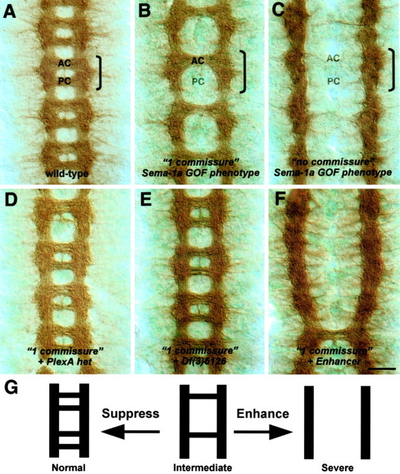
Figure 1.

Dominant enhancer and suppressor screen of a Sema-1a-dependent phenotype. Filleted preparations of stage 14-16 embryos stained with the BP102 antibody to reveal commissural axons are shown. The anterior (AC) and posterior (PC) commissures are labeled in one segment that is outlined by a bracket. Anterior is up. A, In a wild-type embryo, axons cross the midline in the anterior and posterior commissures. B, Ectopic expression of low levels of Sema-1a in midline glia using the Gal4-UAS system in a sema1aP1 mutant background leads to the failure of axons within the posterior commissure to cross the midline (“1 commissure” Sema-1a GOF phenotype = P52-Gal4, UAS:Sema-1a, sema1aP1/sema1aP1). C, Ectopic expression of high levels of Sema-1a in midline glia in a sema1a mutant background enhances the 1 commissure Sema-1a GOF phenotype, resulting in the failure of all commissural axons to cross the midline (“no commissure” Sema-1a GOF phenotype = P52-Gal4, UAS:Sema-1a, sema1aP1/P52-Gal4, UAS:Sema-1a, sema1aP1). D, A deficiency, Df(4)C3, removing one copy of PlexA, the Sema-1a receptor, suppresses the 1 commissure phenotype in B. E, A deficiency, Df(3)5126, removing one copy of Gyc76C, suppresses the 1 commissure phenotype in B. F, A deficiency removing one copy of the genomic region at cytolocation 95F enhances the 1 commissure phenotype in B. G, Summary of Sema-1a GOF commissural axon phenotypes from the dominant enhancer and suppressor screen. Suppression of the intermediate 1 commissure phenotype yields a more wild-type-like architecture, whereas enhancement results in a more severe phenotype in which most axons do not cross the midline. Scale bar: A-F, 10 μm.
