Figure 2.
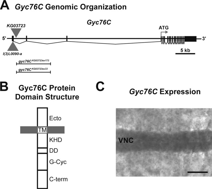
Gyc76C structure and localization. A, Scale representation of the genomic organization of Gyc76C. l(3)L0090-a and KG03723 indicate the locations of P-elements within the Gyc76C gene. Vertical bars and filled boxes represent exons. The extents of the lesions in gyc76CKG03723ex173 and gyc76CKG03723ex33 generated by imprecise excision of the KG03723 transposable element are indicated. B, Protein domain organization of Gyc76C. A putative ligand-binding domain is located at the N terminus (Ecto). A transmembrane (TM) domain anchors the protein in the plasma membrane. The KHD, dimerization (DD), guanylyl cyclase (G-Cyc), and C-terminal (C-term) domains are intracellular. C, A filleted preparation of a wild-type stage 14 embryo hybridized with a cRNA probe specific for a region of Gyc76C. The Gyc76C transcript is broadly distributed but is enriched in cells within the ventral nerve cord (VNC). Scale bar, 35 μm.

