
Figure 4.
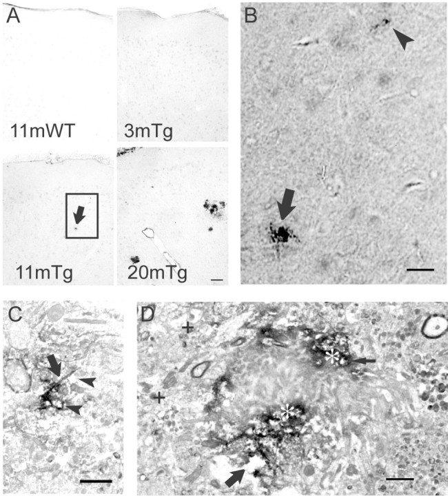
Aβ42 oligomers accumulate in Tg2576 mouse brain. A, Low-magnification light microscopy images of M16 Aβ42 oligomer immunoperoxidase labeling of wild-type (WT) and Tg2576 (Tg) mouse brains at different ages. Aβ42 oligomers were not detected in 11-month-old wild-type (top left) or 3-month-old Tg2576 mouse brains (top right) but were faintly detected as dense micro-deposits (bottom left, arrow) in an 11-month-old Tg2576 mouse. By 20 months, M16 Aβ42 oligomer staining showed increased number and size of microdeposits (bottom right) and also surrounded neuritic plaques. Scale bar, 50 μm. B, High-magnification light microscopic image of the rectangle in A (bottom left), showing a microdeposit (arrow) and even smaller Aβ42 oligomer aggregation before plaques in an 11-month-old Tg2576 mouse (arrowhead). Scale bar, 30 μm. C, Immunoperoxidase EM of Aβ42 oligomer aggregations distant from an obvious plaque. Aβ42 oligomer immunoreactivity appears concentrated along processes (arrow), and structures around the aggregation sites are degenerating (vacuolated spaces, arrowheads). Scale bar, 1μm. D, Ultrastructural localization of Aβ42 oligomers around a plaque. Marked accumulation of immunoperoxidase reaction product (appearing black; asterisks) representing Aβ42 oligomers surrounds an unlabeled plaque core. Areas around the plaque core are degenerated and include degenerating neurites (thin arrow) and abnormal empty spaces (large arrow). For reference, more normal neurites distant from the plaque are also seen (plus signs). Scale bar, 1 μm.