
Figure 5.
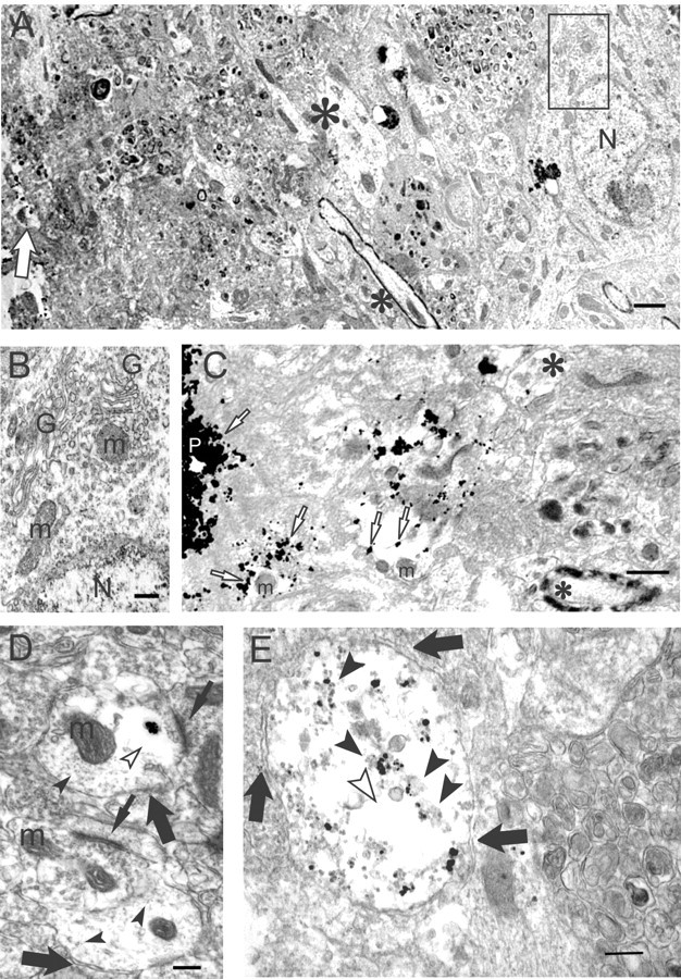
Aβ oligomers by immunogold EM accumulate in dystrophic processes and synapses of Tg2576 mouse brain. A, A lower-magnification image of a degenerated, morphologically abnormal area around a plaque core (left) surrounded by normal-appearing neuropil (right). A degenerating neurite (empty arrow) is observed close to the plaque at the left edge of the figure. Normal dendrite (large asterisk) and a myelinated axon (small asterisk) are at the border of the normal and abnormal areas. A normal neuron is seen distant from the plaque (right); the rectangle within this normal-appearing neuron is enlarged in B, below. N, Nucleus. Scale bar, 1 μm. B, Normal organelles and cytoarchitecture can be seen in this higher-magnification image of the small rectangle within the neuron in A. G, Golgi apparatus; m, mitochondrion; N, nucleus. Scale bar, 250 nm. C, A higher-magnification image of an Aβ oligomer containing degenerating neurites and abnormal empty spaces just around a plaque core (P). Aβ oligomers (empty arrow) are associated with the plaque core, and degenerating neurites are associated with empty spaces. A relatively normal dendrite with an isolated M16 gold particle (large asterisk) and a myelinated axon (small asterisk) are observed distant from the plaque core. m, Mitochondrion. Scale bar, 500 nm. D, A cluster of gold particles representing Aβ42 oligomers in a postsynaptic profile near an active zone of a synapse (top thin arrow). The plasma membranes of two postsynaptic profiles are indicated (large arrows). The cluster of Aβ42 oligomers is surrounded by a clear area (empty arrowhead), in which the normal microtubular network (filled arrowheads) is absent. The lower synaptic profile, lacking Aβ42 oligomers, shows a normal microtubular network throughout. Synaptic active zones are indicated by thin arrows. m, Mitochondrion. Scale bar, 250 nm. E, M16 immunogold EM of a Tg2576 neuritic process. Vertical section through a neuritic process in an 18-month-old Tg2576 mouse brain. Distant from a plaque, substantial Aβ42 oligomer gold particles are associated with granular and fibril-like electron-dense material (arrowheads) in an abnormal pathological-appearing process with poorly defined cytoarchitecture, including empty spaces (empty arrowhead) and lacking any normal organelles, such as mitochondria. Arrows outline the plasma membrane of the neuritic process. Scale bar, 250 nm.