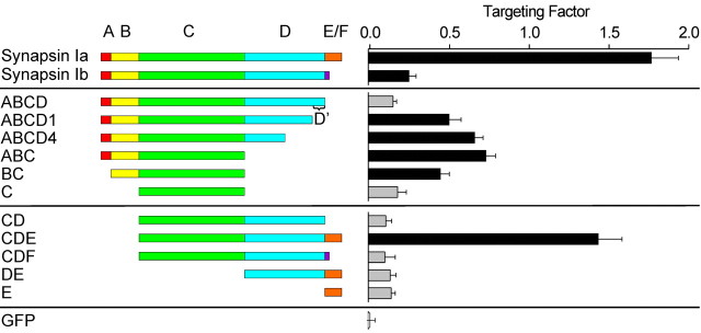
Figure 5.
Targeting of GFP-tagged synapsin I fragments in TKO neurons. Left, Diagram of the synapsin I fragments that were examined. Right, Mean targeting factor determined for each construct expressed in 2-week-old neurons. The targeting factor of soluble GFP was also measured as a control (bottom). Gray bars correspond to fragments that do not target to synapses, as defined by a targeting factor that is statistically indistinguishable from that of GFP. Error bars represent SEM values.

