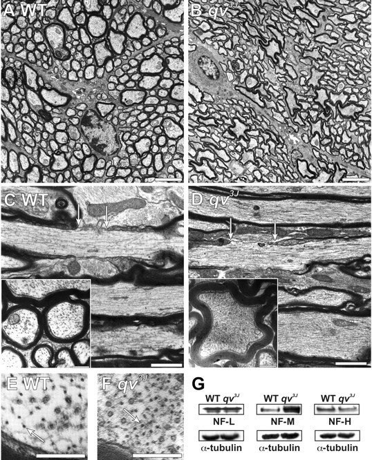
Figure 4.

Four-month-old qv3J mice have dramatic changes in axon shape and cytoskeletal organization. A, B, Transverse optic nerve sections from WT (A) and qv3J (B) mice show that qv3J mouse axons are highly convoluted and not cylindrical. C, D, Longitudinal and transverse (inset) cross sections show that, compared with WT mice (C), qv3J mice have a dramatic increase in the density of cytoskeletal elements (D). Nodes of Ranvier are delineated by arrows. E, F, High magnification of optic nerve cross sections shows qv3J mice (F, arrow) have an increased density of neurofilaments compared with WT mice (E). G, Immunoblotting for neurofilament proteins shows that, in contrast to NF-L and NF-H, the amount of NF-M is increased in qv3J mutant mice. The same blots were probed for α-tubulin as a control for protein loading. Scale bars: A, B, 3 μm; C, D, 1 μm; inset, 0.5 μm; E, F, 0.25 μm.
