Figure 8.
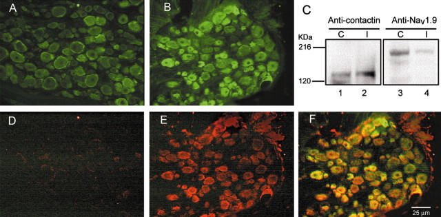
Contactin expression is increased in axotomized DRG neurons. A, In control DRG, contactin is present in most neurons and is peripherally localized. B, In axotomized DRG neurons, there is a substantial increase in contactin immunostaining. C, Western blot analysis shows that transection of the sciatic nerve causes an increase in the contactin protein levels in the DRG. The Western blot was probed with anti-contactin antibody (lanes 1, 2). The increase in contactin protein signal can be seen in ipsilateral (I) DRGs (lane 2) in comparison to contralateral (C) DRGs (lane 1). The blot was then stripped and reprobed with anti-Nav1.9 antibodies (lanes 3, 4). The expected decrease in Nav1.9 protein signal can be seen in ipsilateral DRGs (lane 4) in comparison to contralateral DRGs (lane 3). D, Nav1.3 is not detectable in control DRG neurons. E, There is a substantial increase in Nav1.3 immunofluorescence in DRG neurons (same section as shown in B) after transection of the sciatic nerve. F, Merged images of B and E. Scale bar, 50 μm.

