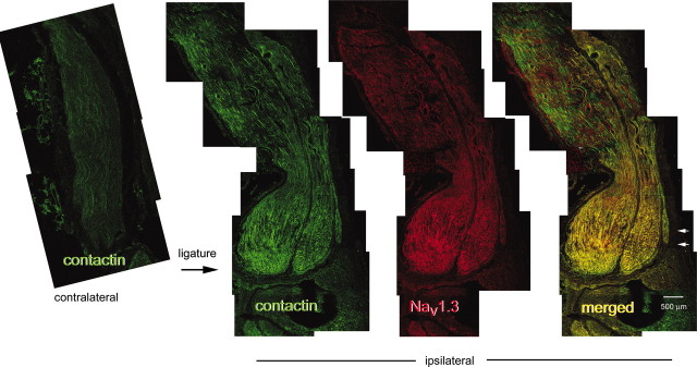
Figure 9.
Nav1.3 and contactin colocalize in sciatic nerve neuroma. Images of uninjured control (contralateral) and transected (ipsilateral) sciatic nerve show that contactin (left) and Nav1.3 (middle) accumulate within axon-like profiles within the neuroma (right, short arrows), with less immunostaining in proximal regions of the transected nerve. Contactin and Nav1.3 exhibit extensive colocalization (right, yellow) within the neuroma. Black arrow “ligature” indicates site of ligation. Scale bar, 500 μm.

