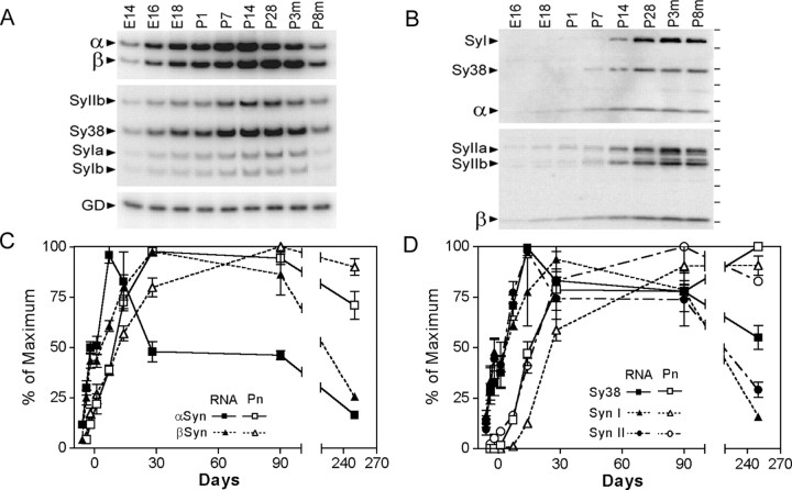
Figure 1.
Post-translational regulation of α-Syn expression in mouse brain with aging. A, Representative autoradiograms of RT-PCR-amplified mRNAs encoding α-Syn (α), β-Syn (β), synaptophysin (Sy38), synapsin I (SyIa, SyIb), synapsin II (SyIIb), and GAPDH (GD). The mRNAs were PCR amplified from cDNAs derived from brains of mouse embryos at embryonic days 14-18 (E14, E16, E18), cortices of postnatal mice at postnatal days 1-28 (P1, P7, P28), and cortices of mature mice at 3 and 8 months of age (p3m, p8m). B, Representative autoradiograms showing the immunoblot analysis of the total SDS-soluble mouse brain extracts for α-Syn, β-Syn, synaptophysin (Sy38), synapsin I (SyI), and synapsin II (SyII). Samples are as indicated in A. C, The levels of α- and β-Syn mRNAs (RNA) and protein (Pn) were quantified from autoradiograms represented in A and B by PhosphorImager analysis and normalized to the maximal level expression achieved. Each value is mean and SEM from three to five independent samples. Quantitative analyses of other presynaptic components are shown in D.

