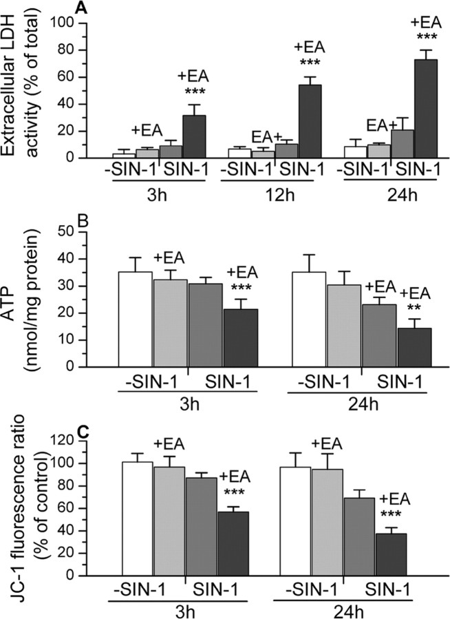
Figure 6.
The effects of treatment with SIN-1 (in HBSS) on LDH release, ATP content, and JC-1 fluorescence in normal astrocytes and astrocytes that were treated with ethacrynic acid (+EA) to deplete mitochondrial glutathione. The responses of these cells to incubation in HBSS without SIN-1 are also presented for comparison. Results for JC-1 fluorescence ratio are expressed relative to values in untreated control cultures (1.4 ± 0.4). The results are shown as mean ± SD. Values significantly different in astrocytes with depleted glutathione exposed to SIN-1 compared with normal cells also treated with SIN-1 are indicated as follows: **p < 0.01 and ***p < 0.001 (one-way ANOVA with Student-Newman-Keuls test). All of the values for the glutathione-depleted astrocytes exposed to SIN-1 were significantly different (p < 0.001) compared with these same cells incubated in HBSS without SIN-1. Significant differences (p < 0.001) were also seen at 24 hr in LDH release, ATP content, and JC-1 fluorescence ratio for normal astrocytes exposed to SIN-1 compared with these cells incubated in HBSS alone. At the earlier time points examined, only the JC-1 fluorescence ratio measured at 3 hr showed a statistically significant difference (p < 0.05) after SIN-1 treatment of these normal astrocytes.

