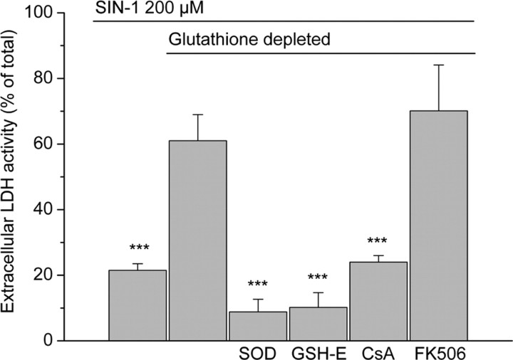
Figure 7.
LDH release at 24 hr after the onset of a 3 hr exposure to 200 μm SIN-1 in cells with mitochondrial glutathione either intact (first bar) or depleted (remaining five bars). Significant protective effects were seen after inclusion of superoxide dismutase (SOD) (500 U/ml; 3 hr), glutathione monoethylester (GSH-E) (5 mm; 30 min), and cyclosporin A (CsA) (2 μm; 3 hr) in the incubation media. Treatment with FK506 (200 nm) was not protective. Values are shown as mean ± SD. ***p < 0.001, compared with SIN-1 exposure in cells with depleted mitochondrial glutathione (one-way ANOVA with Student-Newman-Keuls test; n = 5-10).

