
Figure 8.
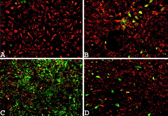
Astrocytes loaded with the mitochondrial membrane potential-sensitive dye JC-1. A, Nontreated cells. B, Normal cells exposed to 200 μm SIN-1 for 3 hr. C, Cells with depleted mitochondria exposed to 200 μm SIN-1 for 3 hr. D, Cells with depleted mitochondria exposed to 200 μm SIN-1 for 3 hr in the presence of 2 μm cyclosporin A. Each picture was taken 24 hr after the onset of the treatment and shows the results of one typical experiment of five. Quantitative analysis of the JC-1 fluorescence revealed that the effect of SIN-1 on cells with depleted mitochondrial glutathione was significantly different when tested in the absence of cyclosporin A (39 ± 9% of control) compared with the presence of this compound (74 ± 6%) (p < 0.01; one-way ANOVA with Student-Newman-Keuls test; n = 5).