Figure 3.
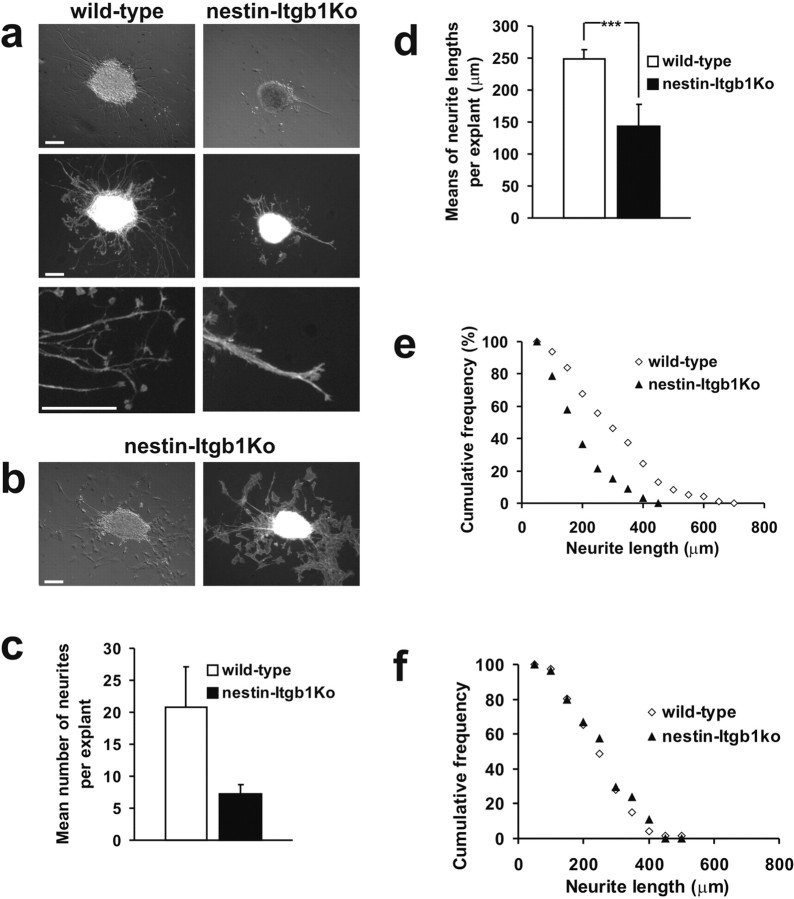
Neurite outgrowth assay in vitro. a, Phase-contrast and fluorescent images of spinal motor neuron explants prepared from E10.5 wild-type and nestin-Itgb1Ko embryos plated on poly-d-lysine/LN-coated coverslips. Wild-type explants formed numerous processes. In explants derived from nestin-Itgb1Ko embryos the neurite outgrowth was strongly impaired, and neurites formed fascicles (bottompanel). b, The growth of β1-deficient motor neurons was not impaired on cocultured fibroblasts. c, The mean number of neurites per explant on LN substrates was reduced significantly in nestin-Itgb1Ko as compared with wild-type embryos. d, The results of the means of neurite lengths per explant on LN substrates were calculated, demonstrating a strong reduction in neurite length between wild-type and nestin-Itgb1Ko explants; ***p = 0.0012. Error bars indicate SEM. The SEM was determined and a Student t test was performed. e, Cumulative frequency distribution plot of neurite length on LN substrates shows a shift for nestin-Itgb1Ko explants toward shorter neurite lengths. f, Cumulative frequency distribution plot of neurite length on fibroblast substrates shows no difference in neurite length between wild-type and nestin-Itgb1Ko explants. Scale bars: a, b, 120 μm.

