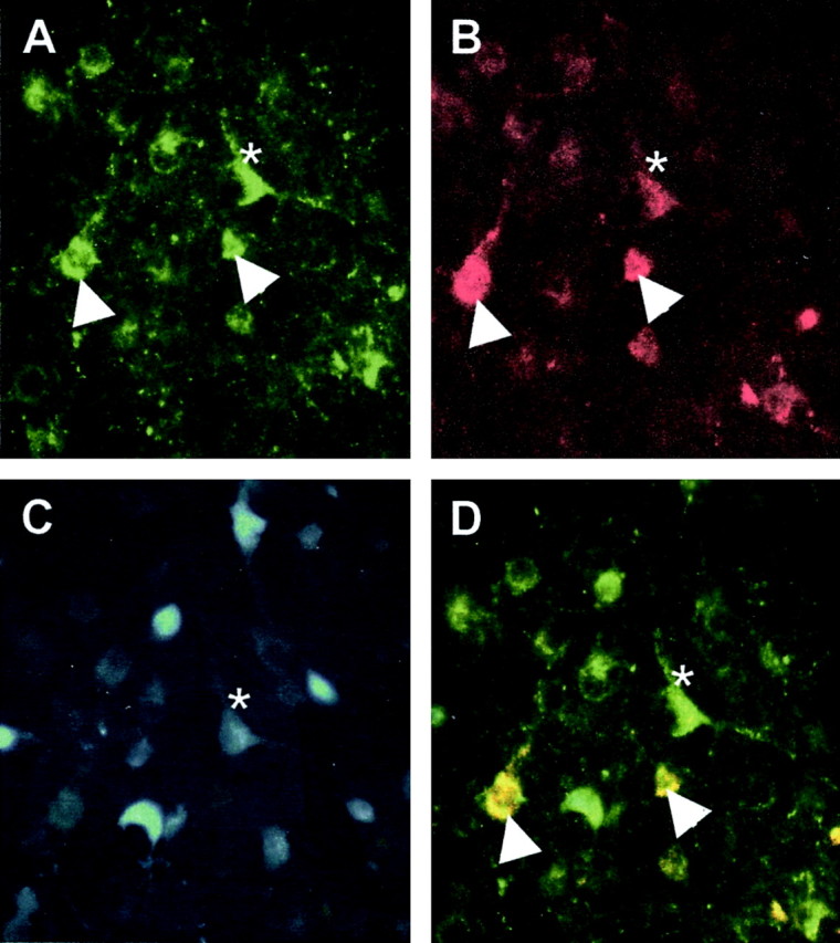
Figure 7.

Photomicrograph of retrogradely labeled cells from the ARH after injection of FG into the MPN. The same field imaged to excite the FITC labeling Y1R (A), tetramethylrhodamine (TRITC) labeling β-END (B), and FG-labeling efferent projections (C) from the MPN. The images are combined in D. Doubly labeled β-END and Y1R immunoreactivity are yellow (arrowheads), and triple-labeled are white (asterisk).
